Structural Organization in Animals
Structural Organization in Animals
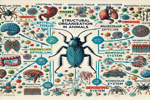
Tissues
Tissues are groups of cells that are similar in structure and function and work together to perform a specific activity. There are four types of tissues in animals:
- Epithelial Tissue
- Covers external surfaces and lines internal cavities and organs.
- Cells are tightly packed with minimal intercellular space and rest on a non-cellular basement membrane.
- Lacks blood vessels and receives nutrients by diffusion from underlying connective tissues.
- Has specialized junctions:
- Tight junctions: Prevent substances from leaking between cells.
- Adhering junctions: Cement neighboring cells together.
- Gap junctions: Allow communication between adjacent cells through cytoplasmic connections.
Classification of Epithelial Tissues:
a) Simple Epithelium (Single-layered):- Squamous epithelium: Flat cells; found in the lining of alveoli and blood vessels.
- Cuboidal epithelium: Cube-shaped cells; found in ducts of glands and nephron tubules.
- Columnar epithelium: Tall and pillar-like cells; found in the lining of the stomach and intestines.
- Pseudostratified epithelium: Appears multilayered but is single-layered; found in the respiratory tract.
b) Compound Epithelium (Multilayered):
- Stratified epithelium: Multiple layers, providing protection; found in skin.
- Transitional epithelium: Specialized for stretching; found in the urinary bladder.
- Muscular Tissue
Specialized for contraction and movement due to the presence of contractile proteins, actin, and myosin.- Striated (Skeletal/Voluntary) Muscle: Multinucleated, cylindrical cells with striations; under voluntary control; attached to bones.
- Unstriated (Smooth/Involuntary) Muscle: Spindle-shaped, uninucleate cells without striations; found in walls of internal organs.
- Cardiac Muscle: Branched, uninucleate cells with faint striations; interconnected by intercalated discs; found in the heart.
- Nervous Tissue
- Specialized for receiving and transmitting impulses.
- The basic unit is the neuron, the longest cell in the body.
- Structure of a neuron:
- Dendrites: Receive signals.
- Cyton (Cell Body): Contains the nucleus.
- Axon: Long projection transmitting impulses; may be covered by a myelinated sheath with Nodes of Ranvier.
- Axon Terminals: Transmit signals to other neurons or muscles.
ORGAN AND ORGAN SYSTEM
Cockroach (Periplaneta americana)
- Phylum: Arthropoda
- Class: Insecta
- Habitat: Nocturnal, omnivorous, found in warm and moist environments.
Morphology
- Body size: 1/4 to 3 inches.
- Covered with a chitinous exoskeleton divided into hardened plates called sclerites (tergites on the dorsal side, sternites on the ventral side), connected by a flexible arthrodial membrane.
- Head: Triangular, with compound eyes and long, segmented antennae.
- Mouthparts:
- Labrum: Upper lip.
- Mandibles: Chewing structures.
- Maxillae: Assist in handling food.
- Labium: Lower lip.
- Hypopharynx: Tongue-like structure.
- Thorax: Divided into prothorax, mesothorax, and metathorax, each with a pair of legs.
- Abdomen: Segmented with spiracles for respiration.
Anatomy
- Digestive System
- Path: Mouth → Oesophagus → Crop (food storage) → Gizzard (grinding food) → Hepatic caeca → Midgut → Hindgut → Rectum → Anus.
- Hepatic caeca: Secrete digestive enzymes.
- Circulatory System
- Open type with haemolymph (blood-like fluid).
- Heart: A tubular structure with ostia (valvular openings) that pump blood anteriorly.
- Respiratory System
- Composed of tracheae, with external openings called spiracles.
- Tracheoles carry oxygen directly to tissues.
- Excretory System
- Malpighian tubules: Excrete nitrogenous wastes as uric acid (uricotelic).
- Other organs: Fat bodies, nephrocytes, and urecose glands.
- Nervous System
- Ganglia: Three pairs in the thorax and six pairs in the abdomen.
- Sense Organs: Antennae, compound eyes, maxillary palps, labial palps, and anal cerci.
- Compound eyes have 2000 ommatidia, providing mosaic vision.
- Reproductive System
- Male: Testes → Vas deferens → Seminal vesicle → Ejaculatory duct.
- Female: Ovaries → Oviduct → Vagina → Genital chamber.
- Fertilized eggs are enclosed in oothecae (9–10 per female).
- Development: Paurometabolous (through nymphal stages), with 13 moults to reach adulthood.
Structural Organization in Animals
Read also –
- NCERT Class 11 Biology Textbook
- Structural Organisation in Animals – BYJU’S
- Structural Organisation in Animals – Toppr
- NCERT Solutions – Learn CBSE
- Structural Organisation in Animals – Vedantu
- ExamFear Notes on Structural Organisation
- https://alisciences.com/anatomy-of-flowering-plants/
- https://alisciences.com/diversity-in-the-living-world/
Hi, I’m Hamid Ali, an MSc in Biotechnology and a passionate Lecturer of Biology with over 11 years of teaching experience. I have dedicated my career to making complex biological concepts accessible and engaging for students and readers alike.
Beyond the classroom, I’m an avid blogger, sharing insights, educational resources, and my love for science to inspire lifelong learning. When I’m not teaching or writing, I enjoy exploring new advancements in biotechnology and contributing to meaningful discussions in the scientific community.
Thank you for visiting my blog! Feel free to connect and explore more of my work.